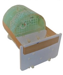
Large ACR MRI Phantom JM Specialty Parts
Description
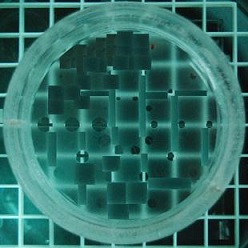
The Large MRI QA Phantom is suitable for scanners capable of all examinations,no matter how many modules are on your application. The Small MRI Phantom is suitable for scanners only capable of extremity exams.
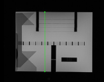
The RIT Radia Diagnostic software automatically analyze MR Images of the above phantoms in seconds. Providing reports on Localizer line length, geometric accuracy, resolution, slice thickness, slice position accuracy, Percent integral uniformity (with full statistics), Percent signal ghosting (with full statistics), Low contrast objects detected with contrast-to-noise reported for each object.
Details
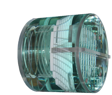
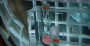 Two cubes with 10 mmol nickel chloride and vegetable fat.
 Four low-density contrast disks with holes of different diameters at 0.002, 0.004, 0.006 and 0.008 inches in thickness. Used to assess MRI scanners' ability to distinguish low contrast objects.
 Low Contrast Detectability: Four sets of plastic membranes with holes 1.5 mm to 7 m in diameter.
Geometric Accuracy, Distortion, Artifacts and Distance Measurements: Array of 10 by 10 square Grid.
Low Contrast Detectability: Four sets of plastic membranes with holes 1.5 mm to 7 m in diameter.
Geometric Accuracy, Distortion, Artifacts and Distance Measurements: Array of 10 by 10 square Grid.
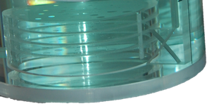
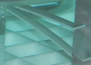 Slice Thickness Accuracy: Counter Descending Wedge.
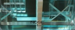 Two sets of paired 45º wedges. Used to precisely measure physical and electronic slice offsets and evaluate small inter-slice gaps.
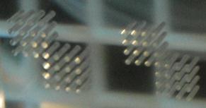 High Contrast Spatial Resolution: Three matrices of holes in an 11 mm thick bar. Hole diameters are 1.1 mm, 1.0 mm and 0.9 mm.
×
![]()
Additional information
Brochure
Example Reports
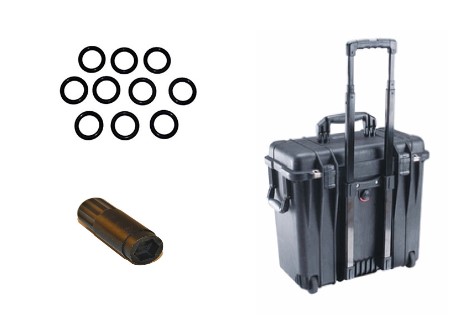
Accessories